Forensics and Jane

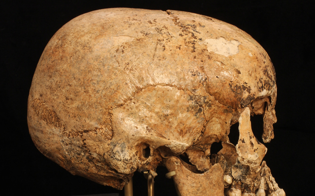
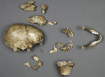
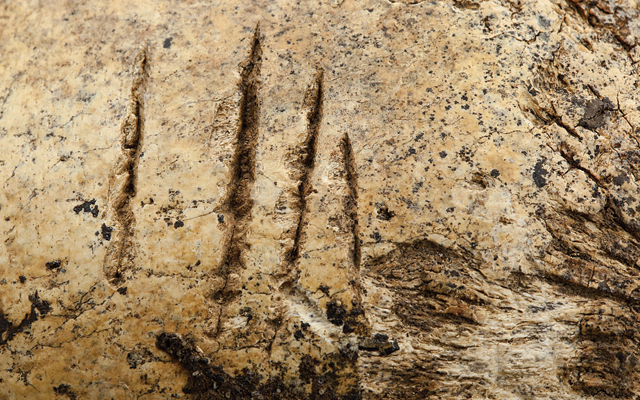
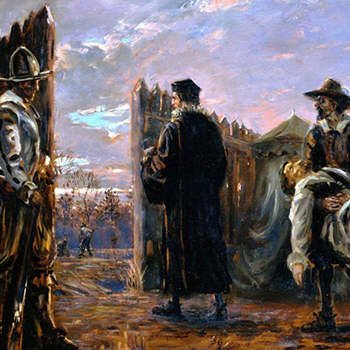
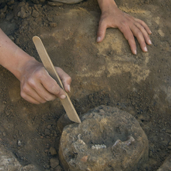
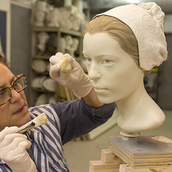
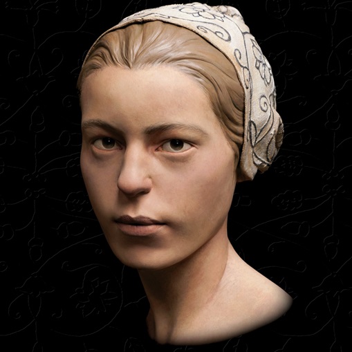
The biological profile of every skeleton is unique, so a bone biography combined with evidence at the scene of excavation can answer many questions about the life of an unidentified person. Over the past 20 years, Dr. Douglas Owsley and his team at the National Museum of Natural History at the Smithsonian Institution have examined several Jamestown skeletons: one that had been shot in the leg and another that proved to be a young boy hit with an arrow in 1607. The Smithsonian provided important clues that another James Fort burial was that of Captain Bartholomew Gosnold, a major planner of the colony. In 2012, Owsley's team was given the challenge of analyzing the remains found in the James Fort L-shaped cellar.
What does forensic science tell us about Jane?
jane-found

The skull of the colonist as it appeared after the pieces found in 2012 were put back together.

Age

Reliable markers for the age can quickly be gained from dental and bone development. Deciduous (baby) teeth are present through childhood and are then replaced with permanent teeth. Around the age of 15, third molars (wisdom teeth) typically begin to erupt from the mandible (jaw). Another maker for age comes from the growth of bones. During growth, bone is expanding, and the area between the bones stays open to allow for that growth. As we age, these fissures close and smooth over. The dentition of this skull has unerupted third molars with partially-formed roots, and the right tibia's growth plate at the knee was just beginning to fuse -- signs that point to an age of 14.

Gender

Cranial shape is an excellent indicator of gender. Females tend to have a more vertical forehead, whereas males tend to have a gently sloping forehead and often a strongly-developed brow ridge. Just behind the mandible (jaw) is a small protruding bone know as the mastoid process -- a muscle attachment for that jaw that is often very developed in males and quite small in females. Near the base of the cranium are a series of muscle attachments known as the nuchal area. Females tend to exhibit less development of nuchal attachments in comparison to males. Two of the largest portions of the cranium are the parietal bones. Located in the back of the skull, these boney plates tend to bulge out ("bossing") as we transform from infant to young adult. Females often exhibit this “parietal bossing” more prominently than males. The size and shape of this skull is small and elongated, with a vertical forehead that is characteristic of an early 17th-century English female.

How does isotopic testing locate Jane's origin?
jane-found

Skeletal remains

Additional, discrete ethnic data about these bones found at James Fort come from isotopic testing. Stored within our bone structure is stable data that contain heavy oxygen, nitrogen, and carbon isotopes. Some of this data is locked in to our skeletal structure as we develop to adulthood, whereas other data accumulates as we consume different types of food. Oxygen isotopes are stored in our teeth in childhood and can be used with other information to determine where a person grew up.

Testing the bone's stable carbon and nitrogen isotopes indicated this young woman was a recent immigrant; the chemical composition of the bone reflects a European, wheat-based diet and not an American, corn-based diet. Jane's isotopic values told the story of a middle- to upper-class individual born along the coastal plains of southern England. Or was she a maidservant eating the same food as the gentry she served?

What does science tell us about Jane and cannibalism?
jane-found

Cuts in the skull fragments indicate the female colonist was a subject of cannibalism.

Simple magnification is one of the most useful tools that a forensic anthropologist has. Magnifying glasses, stereo zoom microscopes, and scanning electron microscopes (SEM) see what the natural eye cannot. Although the form of some microscopes hasn't changed in centuries, the modern technology of automation and digital imaging has transformed microscopes into precise instruments that illuminate unseen evidence.

Under intense magnification, Jane's mandible told a big part of the forensic story. Knife cuts on the jaw showed that the cutting was done in quick sawing motions without a clear pattern -- as if the cutter was tentative and inexperienced. Knife jabs to the bottom of the jaw clearly indicated an intent to remove flesh.

How did Dr. Douglas Owsley and his team at the National Museum of Natural History at the Smithsonian Institution know that post-mortem butchering of the skull and leg is evidence of cannibalism?

Four chops to the middle forehead represent a tentative, failed attempt to open the cranium. Bone in the back of the head shows a series of deep chops; these forceful blows fractured the cranium along its midline. Bone below the right eye socket (maxilla) has a series of small, fine cuts from a knife being used to remove cheek muscles. Numerous small knife cuts and punctures in the mandible reflect attempts to remove tissues from both the inside and outside of the lower jaw. The left temporal bone was punctured by a small, rectangular tool. The narrow tip of the tool caused this compression fracture as it pried the bone from the side of the head to gain access to the brain. The right tibia bone has a chop halfway through its shaft. The blade entered the leg bone below the knee and from behind, breaking the shaft and exposing the marrow. Fine cuts indicate a sharp knife was also used to remove the leg.

How does science help us see Jane?
jane-found

The team at StudioEIS in New York used a 3D model based on digital images to sculpt a lifelike Jane.

Reconstructing a face from the pieces of skull that once supported it is a mix of art and science that can revive an appearance from four centuries ago.

Gaps in the skull where fragments are missing can be filled with computer imaging. A computed tomography or CT scan of each of the skull fragments gives a scientist a digital representation of exactly what one sees if holding the bone in hand, including knife cuts on the bone. Then the scientist can create missing parts of the skull by copying what did survive on the opposite side and then reversing those pieces. With such mirror imaging, a skull can be aligned to approach its original shape.

In that way, Jane's missing right malar bone was replaced, as was her central incisor and the maxilla with teeth on one side. The split up the middle of her skull was repaired. Spaces were closed around her upper arch. Her bones have returned to their proper anatomical position.

A resin cast of Jane's skull could then be made. Guided by scientifically-generated thickness markers on that mold, an experienced forensic sculptor skillfully applied modeling clay. Hairstyle and eye and hair color were based on 17th century portraits.


Jane's History

Jane's History

Archaeology

Archaeology

Exhibit

Exhibit

Jane in the News

Jane in the News